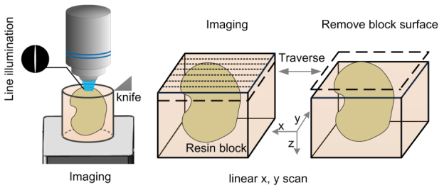
Fig. 7.
Schematic diagram for three-dimensional fluorescent imaging. The resin-embedded biological sample was mounted on a precision motion stage that moves between the stage-scanning microscopy and the microtome. Strip imaging methods combined with line illumination were applied for rapid surface imaging. After the surface layer of specimen was imaged, the recorded layer (1-μm thick) was removed by a fixed diamond knife. The sectioning-imaging cycles was repeated for collecting three-dimensional imaging data sets.

