
Figure 3.
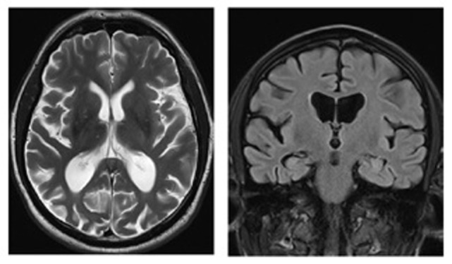
Magnetic resonance images (axial T2, coronal FLAIR) of a patient with posterior cortical atrophy demonstrating marked regional atrophy in the occipitotemporal regions and relative preservation of the hippocampi. Patient under the care of GTP; published with patient’s authorization.