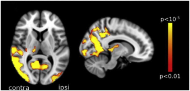
Figure 4.

Voxel-based morphometry analysis comparing a group of posterior cortical atrophy (PCA) patients and age-matched healthy controls. Areas with most significant atrophy (highlighted in red-yellow) in PCA patients included the lateral and anterior occipital cortex, with some loss also noted in the parietal lobe, more marked in the hemisphere contralateral to the visual field defect, here represented on the left. Reproduced from Millington et al. (38), available under the terms of the Creative Commons Attribution License (https://creativecommons.org/licenses/by/4.0/).
