
Figure 8.
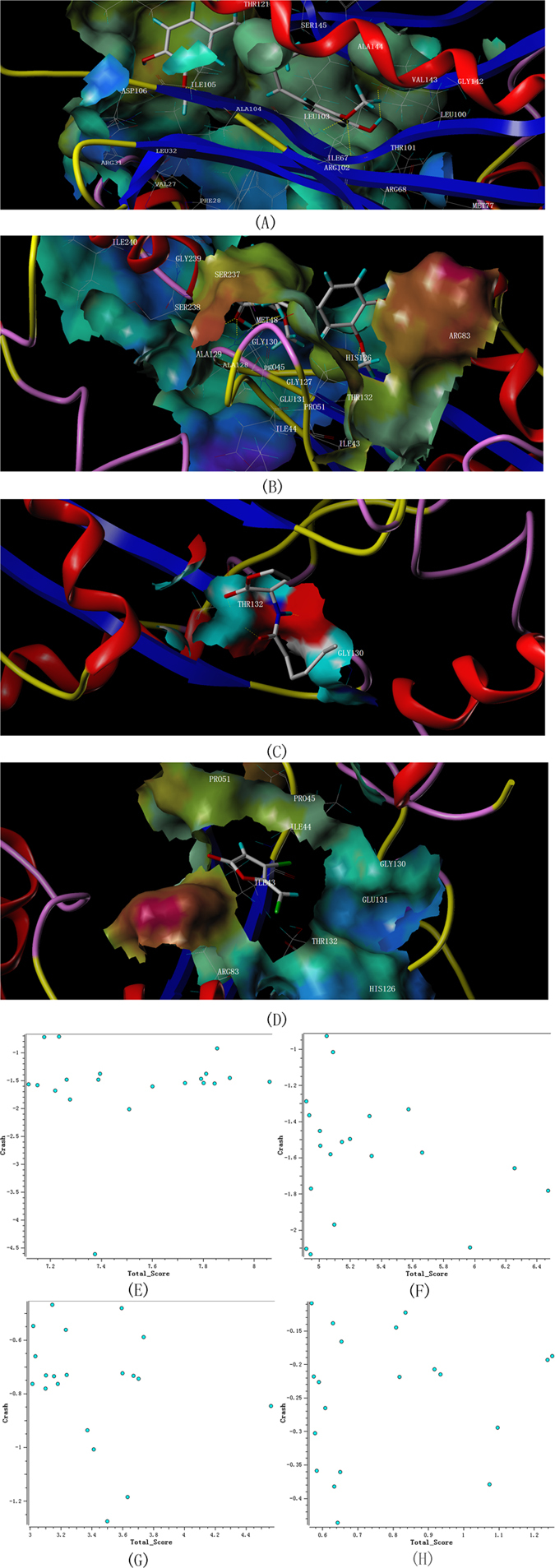
kGraphical representation of LuxI and LuxR type proteins of A. sobria docked with curcumin, C6-HSL and furanone C-30 as well as the conformations of the ligands. Yellow dashed line represents the Hydrogen-bonds. Residues in the active site were labeled. (1) Curcumin docked with modeled LuxI type protein (2) Curcumin docked with modeled LuxR type protein (3) C6-HSL docked with modeled LuxR type protein (4) Furanone C-30 docked with modeled LuxR type protein (5) Conformations of curcumin docked with LuxI type protein (6) Conformations of curcumin docked with LuxR type protein (7) Conformations of C6-HSL docked with LuxR type protein (8) Conformations of furanone C-30 docked with LuxR type protein.