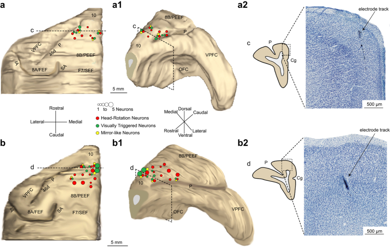
Figure 2.
Histological reconstruction of the recorded regions and functional maps of MK1 and MK2. (a,b) 3-D reconstruction (dorsal view) of the left hemisphere of MK1 (a) and MK2 (b), with superimposed number and class of neurons recorded from each site. (a1,b1) 3-D reconstruction (rostro-lateral view) of the same hemispheres of MK1 (a1) and MK2 (b1). (a2–b2) Examples of coronal sections at high magnification (photomicrograph of Nissl-stained section), showing where electrode tracks were identified. Dashed black lines in (a,b) and dashed black boxes in (a1–b1), labeled “c” and “d” indicate the position from which each coronal section was taken. SA, superior arcuate sulcus; IA, inferior arcuate sulcus; P, principalis sulcus; VPFC, ventral prefrontal cortex; OFC, orbitofrontal cortex.

