
Figure 8.
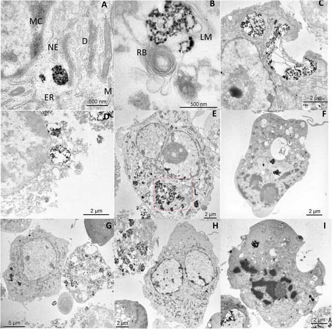
TEM images of BV2 cells containing 100 µg/ml of PAA-MNPs after 30 min of MHT at 46 °C. (A) vesicle-containing MNPs, ER: endoplasmatic reticulum, NE: nuclear envelope, MC: marginal chromatine, (D) dictyosome and M: mitochondrie. (B) MNPs cluster wrapped in lysosomal membrane (LM) and close to a residual body (RB). (C) Disrupted cell membrane near a large PAA-MNPs cluster after MHT. (D) Typical image of a completely damaged cell due to AMF and PAA-MNPs. (E) Cell with lysosome containing PAA-MNPs. Apoptotic cells (F and I) and dead cells (G and H) were found to coexist after MHT with (seemingly) alive cells containing high variable amounts of PAA-MNPs incorporated.