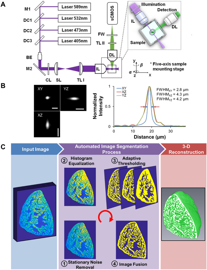
Figure 1.
Light-Sheet Fluorescent Imaging, Histogram Image Processing, and 3-D Reconstruction. (A) Collimated lasers are transmitted through an illumination lens (IL) to generate a light-sheet sectioning the sample. The detection arm includes an objective lens (DL) positioned orthogonally to the illumination path for fluorescence detection. The detection axis needs to exactly conjugate the illuminated plane with the camera CMOS plane. (B) We obtained the lateral and axial optical spatial resolution by measuring the point spread function (PSF) of 0.5 μm beads, expressed as the FWHM from the point source in the XY, XZ, and YZ planes. (C) Summary of the image reconstruction and 3-D rendering process in 3 phases encompassing input image, automated image segmentation, and 3-D reconstruction. BE: beam expander. CL: cylindrical lens. DC: dichroic mirror. DL: detection lens. FW: filter wheel. FWHM: full width at half maximum. IL: illumination lens. M: mirror. sCMOS: scientific complementary metal oxide semiconductor. SL: scan lens. TL: tube lens. Scale bar: 5 μm.

