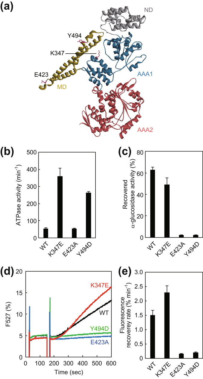
Figure 4.

Characterization of hyperactive and repressed mutants of TClpB. (a) Monomeric structure of TClpB (PDBid:1QVR)21 is shown. The N-domain (ND), M-domain (MD), AAA1, and AAA2 are colored by gray, yellow, blue, and red, respectively. The mutated residues, Lys347, Glu423, and Tyr494 are shown as sticks. (b) ATPase activities of the TClpB mutants were measured. The experimental procedure was the same as in Fig. 2. (c) Disaggregation activities of the TClpB mutants with TDnaK (0.6 μM), TDnaJ (0.2 μM), and TGrpE (0.1 μM dimer) (termed TKJE) were measured by using α-glucosidase as a substrate. The experimental procedure was the same as in Fig. 3a. The time courses (d) and the initial rates (e) of the reactivation of heat-aggregated EYFP by the TClpB mutants with TKJE are shown. The experimental procedure was the same as in Fig. 3b,c. Error bars represent standard deviations of three or more independent measurements. WT, wild-type.
