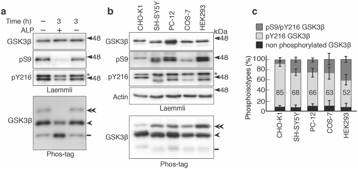
Figure 2.
Phosphoisotypes of endogenous GSK3β in various cell lines. (a) Dephosphorylation of endogenous GSK3β with alkaline phosphatase (ALP). The CHO-K1 cell extract was incubated with 2.5 U/ml ALP for 3 h and was subjected to Laemmli’s (upper) and Phos-tag (lower) SDS-PAGE, followed by immunoblotting with anti-GSK3β, anti-pS9 and anti-pY216 antibodies. In Phos-tag blot the double-arrowhead indicates Ser9/Tyr216 double phosphorylated GSK3β, the arrowhead indicates Tyr216-phosphorylated GSK3β, and the bar indicates nonphosphorylated GSK3β. (b) Immunoblots showing the endogenous GSK3β in cultured cell lines. The cell extracts of CHO-K1, SH-SY5Y, PC-12, COS-7 and HEK293 cells were subjected to Laemmli’s SDS-PAGE, followed by immunoblotting with anti-GSK3β, anti-phospho-Ser9 (pS9) and anti-phospho-Tyr216 (pY216) antibodies. Actin was used as the loading control. An asterisk in the pY216 blot indicates GSK3α. Phos-tag immunoblot of endogenous GSK3β with anti- GSK3β is shown in the bottom panel. The labeling of three bands in Phos-tag is as in (a). (c) Quantification of the GSK3β phosphoisotypes: the upper dark gray section represents phospho-Ser9/phospho-Tyr216, the middle light gray section represents phospho-Tyr216, and the lower black section represents nonphosphorylated GSK3β. The percentages of phospho-Tyr216 are indicated (means ± s.e.m. n = 3). Uncropped immunoblots after Laemmli’s and Phos-tag SDS-PAGE are provided in Supplementary Fig. 6.

