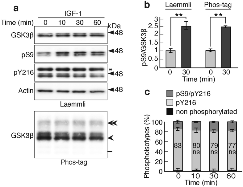
Figure 6.
Effect of IGF-1 on GSK3β activity in cultured primary neurons. (a) Effect of IGF-1 on Ser9 phosphorylation of GSK3β in mouse cortical neurons. Neurons in culture at 5 DIV were treated with 100 ng/ml IGF-1 for the indicated times, and the extracts were subjected to Laemmli’s SDS-PAGE, followed by immunoblotting with an anti-GSK3β, anti-phospho-Ser9 and anti-phospho-Tyr216 or Phos-tag SDS-PAGE, followed by immunoblotting with the anti-GSK3β. Actin is the loading control. An asterisk in the pY216 indicates GSK3α. Double arrowhead, Ser9/Tyr216-phosphorylated GSK3β; arrowhead, Tyr216-phosphorylated GSK3β; bar, nonphosphorylated GSK3β. (b) Levels of pSer9 in neurons that were treated with IGF-1 for 30 min. The ratio of pS9/GSK3β was measured and expressed to that of the control at 0 min. The measurements by Laemmli’s and Phos-tag SDS-PAGE are shown in left and right, respectively. (c) The quantification of three phosphoisotypes of GSK3β in a Phos-tag blot: dark gray, phospho-Ser9/phospho-Tyr216; light gray, phospho-Tyr216; black, nonphosphorylated GSK3β. The percentages of phospho-Tyr216 are indicated (means ± s.e.m. n = 6, ns, not significant). Uncropped immunoblots of GSK3β, pS9, pY216 and actin after Laemmli’s SDS-PAGE and GSK3β after Phos-tag SDS-PAGE are provided in Supplementary Fig. 9b.

