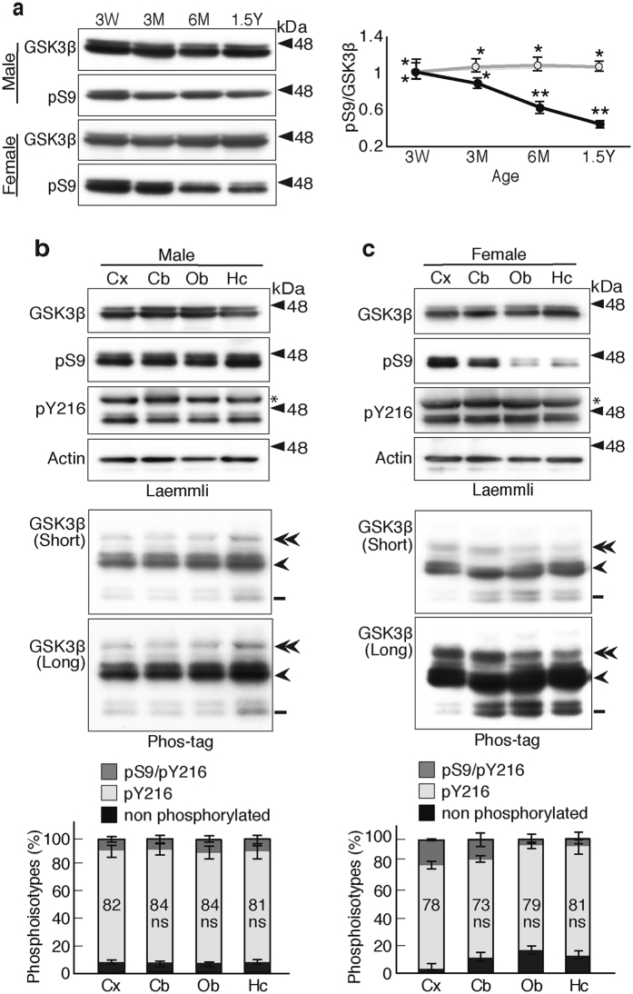
Figure 7.
GSK3β phosphoisotypes in mouse brains. (a) Expression and Ser9 phosphorylation of GSK3β in mouse brain with aging. Extracts of hippocampus of male and female mouse at ages 3 weeks (3 W), 3 and 6 months (3 M and 6 M), and 1.5 years (1.5Y), were immunoblotted with anti-GSK3β or anti-phospho-Ser9 after Laemmli’s SDS-PAGE. The ratio of phospho-Ser9 to total GSK3β was quantified and expressed to the ratio at 3 weeks. Gray represents male and black represents female (means ± s.e.m. n = 6, *p < 0.05, **p < 0.01, one-way ANOVA). (b and c) Analysis of GSK3β phosphorylation in different regions of brains prepared from 1.5-year-old mice; cerebral cortex (Cx), cerebellum (Cb), olfactory bulb (Ob) and hippocampus (Hc). (b) shows the male mice and (c) shows the female mice. The top four panels show Laemmli’s SDS-PAGE and the lower two panels (upper, shorter exposure; lower, longer exposure) show Phos-tag SDS-PAGE that had been immunoblotted with the indicated antibodies. The percentages of three phosphoisotypes of GSK3β are shown below the blots. Dark gray represents phospho-Ser9 and phospho-Tyr216 GSK3β, light gray represents phospho-Tyr216, and black represents nonphosphorylated GSK3β. The percentages of phospho-Tyr216 are indicated (means ± s.e.m. n = 6, ns, not significant). Uncropped immunoblots of GSK3β, pS9, pY216 and actin after Laemmli’s SDS-PAGE and GSK3β after Phos-tag SDS-PAGE are provided in Supplementary Fig. 10.

