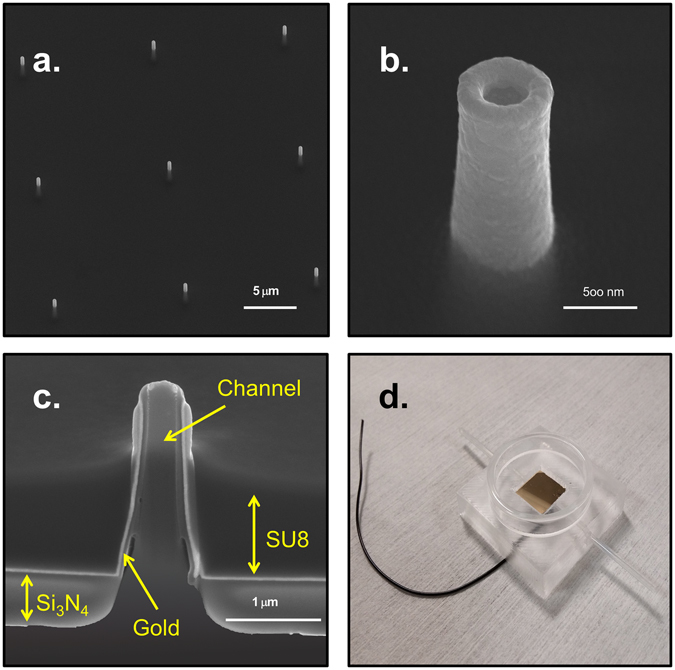
Figure 2.

(a–c) SEM images of the 3D hollow nanoelectrodes embedded in the epoxy polymer SU8. Respectively, 3 × 3 array of nanoelectrodes, single nanoelectrode and cross section of a single hollow nanoelectrode. The different layers are indicated by the yellow arrows. (d) Top view of the PDMS microfluidic chamber. The wire is coming out from the PDMS being bonded by silver paste with the gold spattered onto the device making all the nanoelectrodes connected together. The glass ring around the device allows the cell culture on to the nanofluidic electrodes.
