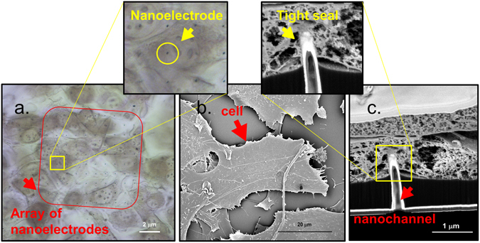
Figure 3.

(a) Transmission image of NIH-3T3 cells fixed on an array of hollow nanoelectrodes – the black dots inside the red square – embedded in SU8. (b) SEM top view image of CPD dried NIH-3T3 cells cultured on top of the hollow nanoelectrodes. (c) Cross section of CPD dried cell grew on top of a hollow nanoelectrode. From this image it is possible to see all the layers of the fabrication (Si3N4 membrane 100 μm thick, SU8) and the hollow 3D nanoelectrode that is engulfed by the cellular membrane.
