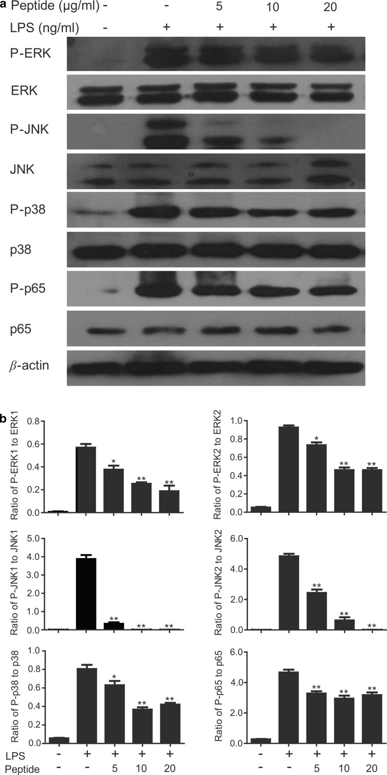
Fig. 9.
Effects of cathelicidin-PP on LPS-induced inflammatory response pathways. a Western blot of phosphorylation of ERK, JNK, p38, and NF-κB p65 in peritoneal macrophages. The cells were incubated with LPS (100 ng/ml) and different concentrations of cathelicidin-PP (0, 5, 10, and 20 μg/ml). After incubation for 30 min, the cells were collected, and the cytoplasmic or nuclear proteins were extracted for Western blot analysis. b Ratio of P-ERK1 (44 kDa), P-ERK2 (42 kDa), P-JNK1 (54 kDa), P-JNK2 (46 kDa), P-p38, and P-p65 to β-actin. Band densities were analyzed using Quantity One software (Bio-Rad, Richmond, CA, USA). Data were presented as mean ± SEM. *p < 0.05, **p < 0.01, ratios of peptide-treated groups are significantly different from that induced by 100 ng/ml LPS alone

