
Figure 6.
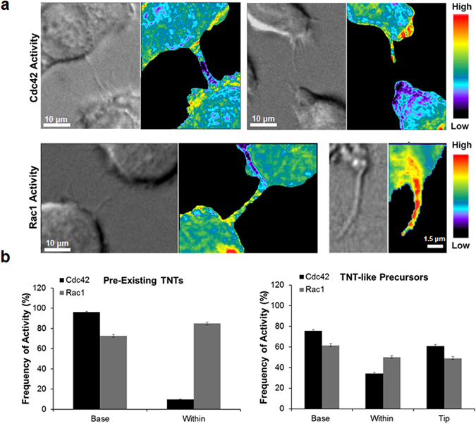
Cdc42 and Rac1 activity during TNT formation in RAW/LR5 macrophages. (a) Representative images of time-lapse ratiometric imaging of RAW/LR5 macrophage cell line with inducible expression of Cdc42 biosensor in pre-existing TNTs (left panels) or TNT-precursors (right panels). Scale bars = 10 µm. (b) Representative images of time-lapse ratiometric imaging of RAW/LR5 macrophage cell line with inducible expression of Rac1 biosensor in pre-existing TNTs (left panels) or TNT-like precursors (right panels). Scale bars: Left = 10 µm, Right = 1.5 µm. (c) Quantitation of the frequency of Cdc42 activity (black bars) or Rac1 activity (grey bars) in the base and within pre-existing TNTs (left graph) or at the base, within and at the tip of growing TNT-like precursors (right graph). Data is represented as an average of at least 20 TNT structures. Error bars represent +/−SEM.