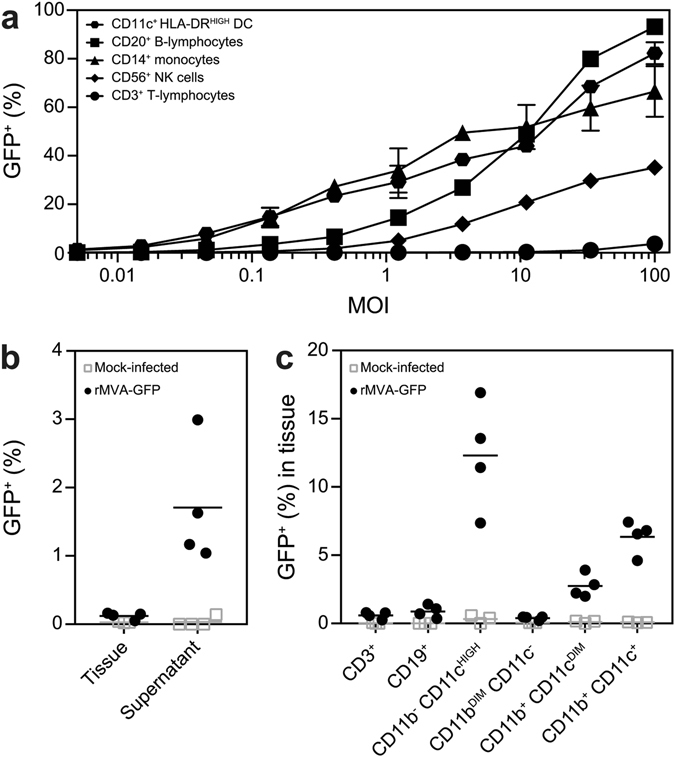
Figure 1.

Populations infected by rMVA-GFP in vitro in human PBMC and ex vivo in mouse lung slices. (a) Human PBMC were inoculated with rMVA-GFP at various MOI. Percentage of GFP+ live cells within DC, B-lymphocyte, monocyte, NK cell and T-lymphocyte populations were determined by flow cytometry at 24 h post-infection. Mean of duplicates and standard deviation are indicated. (b) Lung slices were inoculated with rMVA-GFP and analysed by flow cytometry after 24 h. GFP+ cells in single cell suspensions of lung tissue and culture supernatant were detected. (c) GFP+ cells in single cell suspensions of lung slices were phenotyped by flow cytometry. Populations were defined as CD3+, CD19+, CD11b− CD11cHIGH, CD11bDIM CD11c−, CD11b+ CD11cDIM or CD11b+ CD11c+. Mean percentage of infection per population of four lung slice cultures is indicated.
