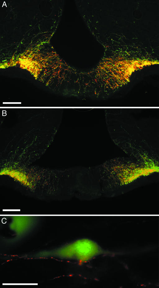
Fig. 4.
Coronal sections through the medial region of the ME of EGFP+/GnRH1 rats immunoreactive to GnRH subtypes are shown. (A and B) Cy3-labeled (red) fibers immunoreactive to GnRH1 (LRH 13) (A) or GnRH2 (aCII6) (B). EGFP+ fibers coexpressing either nonmammalian GnRH appear in yellow. (C) Cy3-labeled GnRH2 (aCII6) fibers (red) in close contact with EGFP+/GnRH1 soma (green) in the septal–preoptic area are shown. (Scale bars: A and B, 100 μm; C, 50 μm.)

