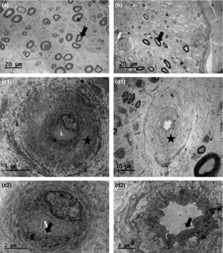
Figure 2.

Sural nerve of a patient with type 2 diabetes at baseline (a), who was severely affected by neuropathy at follow‐up (b; arrows indicate myelinated axons). The microvessels of a control subject (c1; higher magnification in c2) and a patient with type 2 diabetes (d1; higher magnification in d2) are shown. Asterisks indicate basement membrane (c1 and d1) and arrows indicate endothelium (c2 and d2)
