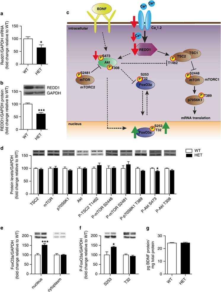
Figure 3.
Cacna1c heterozygous mice have lower REDD1 mRNA and protein levels as well as altered Akt and FoxO3a protein in the PFC. (a, b) Cacna1c HET mice have significantly lower REDD1 mRNA (a) and protein (b) in the PFC compared with WT littermates (mRNA: WT n=9, HET n=11; protein: WT n=7; HET n=6). (c) Schematic representation of the altered signaling pathway in the PFC of HET mice. Red and green arrows depict changes seen basally in cacna1c HET mice. Solid arrows connecting signaling proteins represent pathways shown in the brain. Dashed arrows indicate pathways shown in other systems. (d) Cacna1c HET mice have decreased protein levels of phospho P-Akt S473 in the PFC compared with WT mice (WT n=7, HET n=6). (e) Cacna1c HET mice have significantly higher protein levels of FoxO3a in the nucleus but not the cytoplasm of the PFC, compared with WT mice (WT n=6, HET n=6). (f) Cacna1c HET mice have significantly higher levels of P-FoxO3a S253 but not T32 in the nucleus of the PFC compared to WT mice (WT n=6, HET n=6). (g) Cacna1c HET mice show no difference in BDNF protein levels in the PFC compared with WT mice (WT n=10, HET n=10). *p<0.05, ***p<0.001 vs WT. Error bars are mean±SEM.

