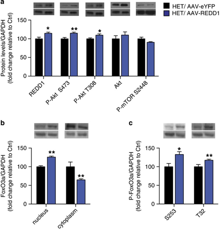
Figure 5.
REDD1 over-expression in PFC of cacna1c heterozygous mice increases levels of phosphorylated Akt and nuclear FoxO3a in the PFC. (a) AAV-REDD1 in the PFC of cacna1c HET mice significantly increases protein levels of REDD1 and P-Akt at S473 and T308 compared with control AAV-eYFP HET mice. (b) AAV-REDD1 in the PFC of cacna1c HET mice significantly increased FoxO3a protein in the nucleus and decreased levels in the cytoplasm compared with control AAV-eYFP HET mice. (c) AAV-REDD1 in the PFC of cacna1c HET mice significantly increased levels of P-FoxO3a protein at S253 and T32 in the nucleus compared with control AAV-eYFP HET mice (HET/AAV-eYFP n=4, HET/AAV-REDD1 n=5–7). *p<0.05, **p<0.01 vs HET/AAV-eYFP. Error bars are mean±SEM.

