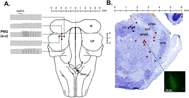
Figure 1.

Injections of the glutamate receptor AMPA were used to functionally identify the “tachypneic area” in the parabrachial nucleus (tPBN). A: Dorsal brainstem image with representative records of the time-averaged phrenic neurogram (PNG, a.u.=arbitrary units). AMPA injection (solid bar) caused marked tachypnea in the tPBN (red circle), but little or no change in four surrounding sites 0.5 mm off-target (solid circles) in an individual adult rabbit. B: Location of fluorescent tracer or Chicago sky blue (700nl) injections into the LC (solid black squares), tPBN (red circles), and KFN (solid black circles) in Nissl stained tissue (Control Study). The inset depicts an example of the tracer spread which has been contrast enhanced to highlight the injection site. LC: locus coeruleus, LPBN: lateral parabrachial nucleus, MPBN: medial parabrachial nucleus, SCM: superior cerebellar peduncle, KFN: Kölliker Fuse nucleus.
