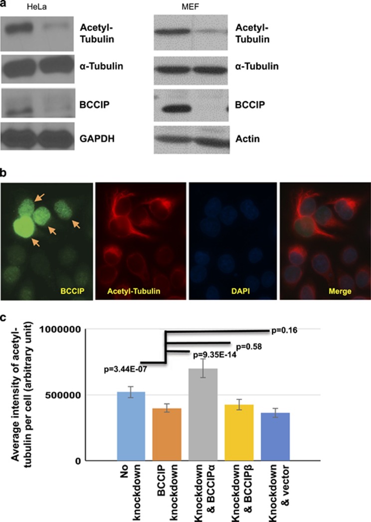
Figure 5.
BCCIP deficiency decreases levels of stable, acetylated microtubules. (a) BCCIP knockdown reduces microtubule acetylation. Control and BCCIP knockdown HeLa cells or MEF control and MEF BCCIP knockout cells were lysed and blotted for the stable microtubule marker, acetyl-tubulin. (b) Levels of acetylated-tubulin in control and BCCIP knockdown cells. HeLa cells expressing BCCIP-shRNA (weak green, un-arrowed) were mixed with control cells (strong green, arrowed), and stained for BCCIP and acetyl-tubulin. (c) The reduced acetyl-tubulin in BCCIP-deficient cell can be rescued by RNAi-resistant BCCIPα. RNAi-resistant YFP-BCCIPα, YFP-BCCIPβ and YFP were re-expressed in BCCIP knockdown cells. These YFP-positive cells were mixed with non-transfected knockdown or control cells (YFP-negative cells). Total fluorescent intensity of acetyl-tubulin in BCCIP knockdown (YFP-negative) and BCCIP re-expressed (YFP-positive) cells on the same staining slide was quantified. Shown are relative intensities of acetyl-tubulin in wild-type cells (first column from left), BCCIP knockdown cells (second column from left) and the knockdown cells expressing different YFP-tagged proteins. Representative images of rescue can be viewed in Supplementary Figure S4.

