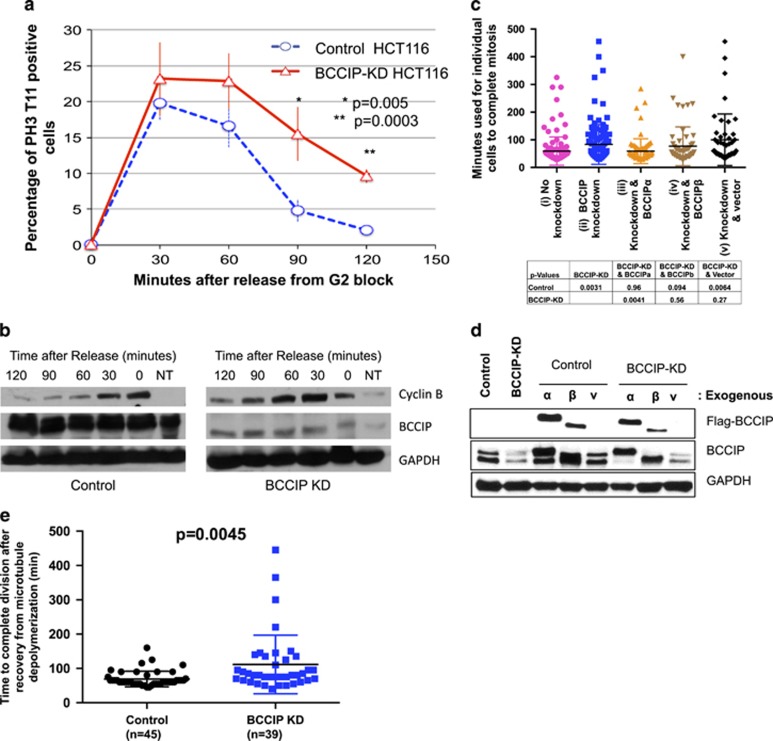
Figure 9.
Delayed mitotic completion in BCCIP-deficient cells. (a, b) BCCIP loss delays anaphase onset. HCT116 cells were blocked at the G2/M boundary by RO-3306 treatment, released and immunostained with pH3-T11. (a) shows the percentages of mitotic cells at different times after the release from the G2/M block. (b) depicts a separate set of experiments where cells were blocked in M phase with nocodazole, released and subjected to western blot at the indicated time points. NT: untreated asynchronized cells used as a loading reference. (c, d) Delayed completion of mitosis in BCCIP-deficient cells. U2OS cells were filmed overnight in an incubated live cell microscope. Frames were acquired every 5 min, and the length of mitosis was quantified from the onset of nuclear envelope breakdown to the formation of a visible mid-body. Shown in (c) is mitotic time in control (i), BCCIP knockdown (ii), and knockdown cells rescued with RNAi-resistant BCCIPα (iii) or BCCIPβ (iv) or control vector (v). The P-values of t-test between the indicated cells are shown. Verification of the re-expression of exogenous BCCIP in the BCCIP knockdown cells is shown in (d). (e) BCCIP deficiency synergizes with spindle poison to delay in mitotic completion. Control and BCCIP knockdown HT1080 cells were treated with nocodazole over ice to completely depolymerize the mitotic spindle. The cells were then washed rapidly three times with warm media and filmed until mitosis was completed.

