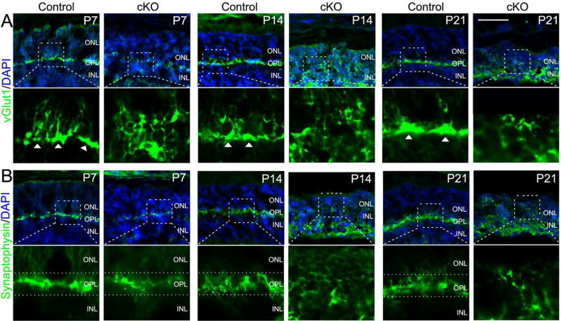
Figure 2. Defective synapse formation in Top2b cKO photoreceptor cells.
Retinal sections from the control and Top2b cKO mice were stained with the synaptic vesicular glutamate transporter marker vGlut1 (A) and synaptic vesicle marker Synaptophysin (B). Arrowheads in (A) indicate the synaptic ends of photoreceptor cells. Dotted lines in (B) indicate the OPL. Boxed areas are shown in a higher magnification. ONL, outer nuclear layer; OPL, outer plexiform layer; INL, inner nuclear layer. Scale bar = 50μm.

