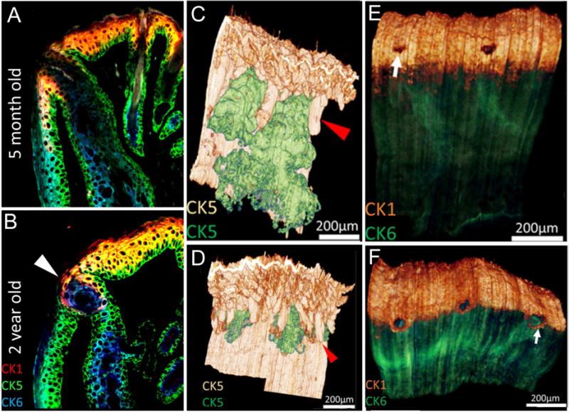
Figure 4.

IT of eyelids from 5 month-old (A, C, E) and 2 year-old (B, D, F) mice. In single sections (A and B) sequentially stained for cytokeratin 1 (CK1, red), 5 (CK5, green) and 6 (CK6, blue) the mucocutaneous junction is detected by the transition from fully keratinized skin (CK1+) to non-keratinized conjunctiva (CK6+). Note the presence of ductal plugging with CK6+ epithelial cells in the 2 year-old eyelid (arrowhead). Volume reconstruction of anti-CK5 staining of eyelids (C and D) with segmentation of the meibomian glands (green) from skin epithelium (gold) shows marked atrophy of the meibomian glands in the 2 year-old compared to the 5 month-old eyelid. Reconstruction of young and old eyelids (E and F) stained for CK1 epidermis (gold) and CK6 conjunctiva (green) show the anterior migration of the mucocutaneous junction (E and F, arrow).
