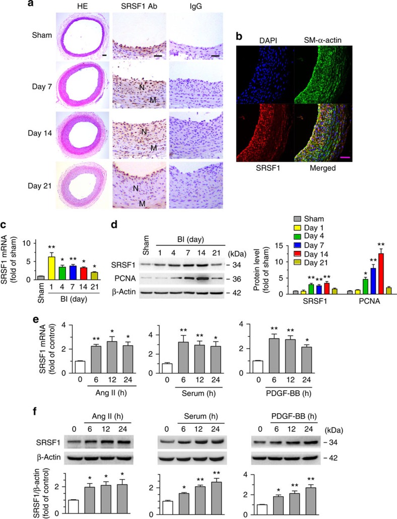
Figure 1. Increased SRSF1 expression in proliferating VSMCs in vivo and in vitro.
(a) Photomicrographs of haematoxylin/eosin-stained carotid arteries from sham-operated and balloon-injured rats. Immunohistochemical staining of vessels with specific anti-SRSF1 antibody revealed SRSF1 mainly in the neointima (N, neointima; M, media). Normal rabbit IgG served as a negative control. Scale bars, 50 μm. (b) Immunofluorescence double staining of injured carotid arteries with specific antibodies against SM-α-actin (green) or SRSF1 (red). Nuclei were stained with DAPI (blue), and yellow indicates their co-localization in the merged images. Scale bars, 50 μm. (c) Real-time PCR showing the mRNA levels of SRSF1 in carotid arteries at 1, 4, 7, 14 and 21 days after balloon injury; n=8 per group. (d) Representative western blots and averaged data showing SRSF1 and PCNA levels in rat carotid arteries at 1, 4, 7, 14 and 21 days after balloon injury; n=8 per group. (e) Real-time PCR data showing the mRNA levels of SRSF1 in HASMCs treated with Ang II (200 nM), serum (10% FBS) or PDGF-BB (10 μg l−1) at 6, 12 and 24 h; n=8 per group. (f) Representative western blots and averaged data showing SRSF1 levels in HASMCs treated as in e; n=7 per group. *P<0.05, **P<0.01, one-way ANOVA (c–f). Data are mean±s.e.m. of five independent experiments (c–f).

