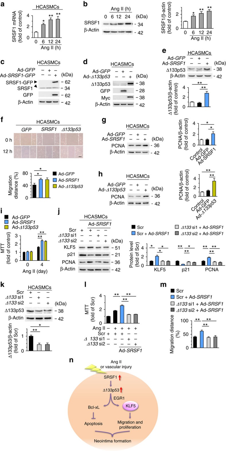
Figure 10. SRSF1 enhances human coronary artery SMC migration and proliferation.
(a) The mRNA levels of SRSF1 in human coronary artery smooth muscle cells (HCASMCs) treated with Ang II at 6, 12 and 24 h; n=8 each. (b) SRSF1 levels in HCASMCs treated as in a; n=9 each. (c) SRSF1 expression in cultured HCASMCs infected with Ad-GFP or Ad-SRSF1-GFP; n=5 each. (d) Δ133p53 expression in cultured HCASMCs infected with Ad-GFP or Ad-Δ133p53; n=5 each. (e) Δ133p53 levels in HCAECs infected with Ad-GFP or Ad-SRSF1; n=6 each. (f) Representative images and averaged data from wound-healing assays in HCASMCs infected with Ad-GFP, Ad-SRSF1 or Ad-Δ133p53 and treated with Ang II; n=9 each. Scale bar, 200 μm. (g,h) PCNA expression in cultured HCAECs infected with Ad-SRSF1 (g) or Ad-Δ133p53 (h); n=5 each. (i) MTT assays of HCASMCs infected with Ad-GFP, Ad-SRSF1 or Ad-Δ133p53 (m.o.i. 100, 48 h) after Ang II for 4 days; n=12 each. (j) KLF5, p21 and PCNA levels in HCASMCs infected with Δ133p53 siRNAs in the presence or absence of Ad-SRSF1 overexpression; n=7 each. (k) Δ133p53 levels in HCASMCs infected with Δ133p53 siRNAs (Δ133 si1 and Δ133 si2); n=7 each. (l) MTT assays showing the SRSF1-mediated enhancement of proliferation was inhibited by Δ133p53 knockdown in HCASMCs; n=15 each. (m) Averaged data from wound-healing assays in HCASMCs infected with Δ133p53 siRNAs with or without Ad-SRSF1 overexpression and treated with Ang II; n=5 each. (n) Schematic outline of SRSF1 -D133p53 -KLF5 axis in VSMC. Ang II, angiotensin II; SRSF1, serine/arginine-rich splicing factor 1; EGR1, early-growth-response gene 1; KLF5, Krüppel-like factor 5; VSMC, vascular smooth muscle cell. Scr indicates scrambled siRNA control. All adenoviral infection above is 100 m.o.i. for 48 h. Concentration of Ang II is 200 nM. Scr indicates scrambled siRNA control. *P<0.05, **P<0.01, one-way ANOVA (a,b,e–m). Data are mean±s.e.m. of four independent experiments (a,b,e–m).

