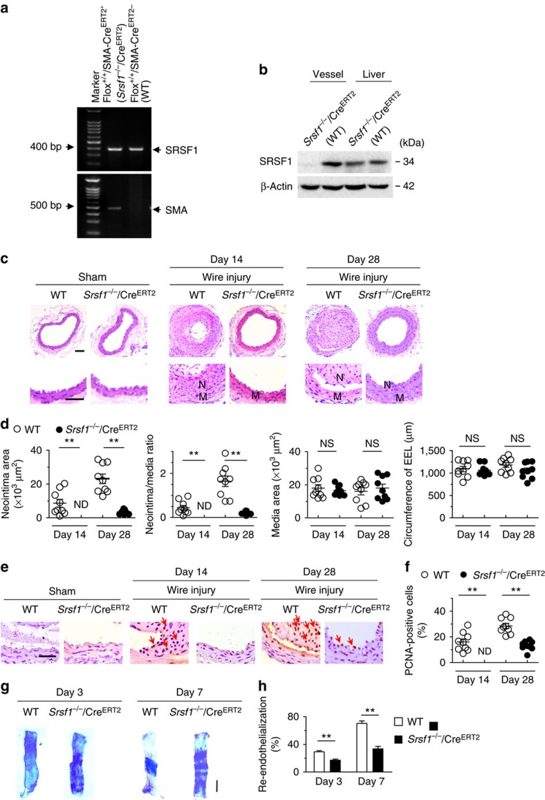
Figure 3. Inducible SMC-specific SRSF1 deficiency inhibits neointima formation.
(a) PCR showing genotyping of WT (Flox+/+CreERT2−) and Srsf1−/−/CreERT2 (Flox+/+CreERT2+) mice. Band in the upper image indicates the product from SRSF1flox/flox, and lower image indicates product from SMA-CreERT2. (b) Representative western blots showing SRSF1 levels in the vessel and liver from inducible smooth muscle cell-specific Srsf1 knockout (Srsf1−/−/CreERT2) mice. (c,d) Representative photomicrographs of haematoxylin and eosin staining (scale bars, 50 μm) (c) and averaged data (d) of the neointimal area, neointima/media ratio, media area and circumference of external elastic lamina (EEL) of carotid arteries from Srsf1−/−/CreERT2 and WT control mice 14 and 28 days after wire injury (N, neointima; M, media); n=9 per group. (e,f) Representative photomicrographs of immunohistochemical staining (scale bar, 25 μm) (e) and averaged data (f) showing the percentages of PCNA-positive cells in carotid arteries from Srsf1−/−/CreERT2 and WT control mice 14 and 28 days after wire injury. Arrows indicate PCNA-positive cells (dark brown); n=9 per group. (g,h) Representative pictures (g) and averaged data (h) of re-endothelialization. Re-endothelialization was quantified in Evans blue-stained carotid arteries at 3 and 7 days after vascular injury. Blue staining indicates endothelial denudation. Scale bar, 1 mm; n=9 per group. **P<0.01, NS, not significant; Student’s t-test (d,f,h). Data are mean±s.e.m. of five independent experiments (d,f,h).

