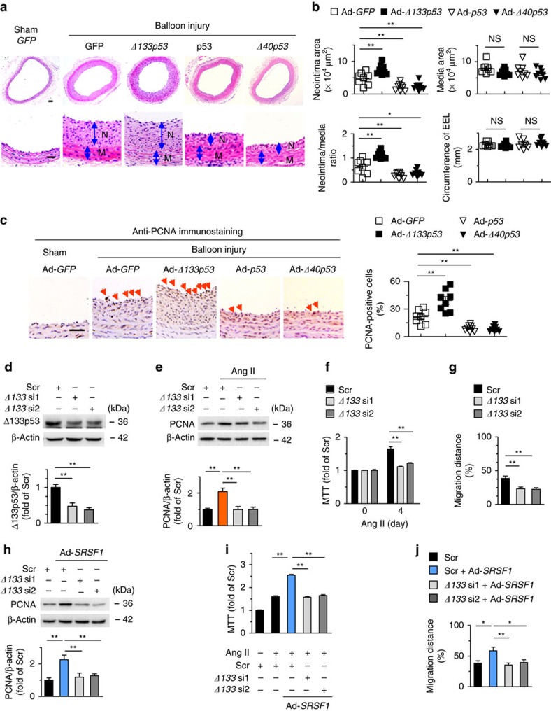
Figure 6. Δ133p53 promotes neointima formation after vascular injury.
(a,b) Hematoxylin/eosin staining (a) and averaged data (b) of the neointimal area, neointima/media ratio, media area, and circumference of external elastic lamina (EEL) of rat carotid arteries transfected with indicated adenovirus 2 weeks postinjury (N, neointima; M, media); n=8 each; scale bar, 50 μm. (c) PCNA staining and PCNA-positive cells in rat carotid arteries treated as in (a) 2 weeks postinjury. Arrows indicate PCNA-positive cells; n=8 each; scale bar, 25 μm. (d) Δ133p53 siRNAs (Δ133 si1 and Δ133 si2) knock down Δ133p53 level in HASMCs; n=8. (e–g) PCNA level (e), MTT assays (f) and wound-healing assays (g) in HASMCs infected with Δ133p53 siRNAs after Ang II stimulation; (e), n=8; (f), n=11; (g), n=9. (h) PCNA levels in HASMCs infected with Δ133p53 siRNAs in the presence or absence of Ad-SRSF1; n=6. (i) MTT assays showing SRSF1-mediated enhancement of proliferation was inhibited by Δ133p53 knockdown; n=12. (j) Averaged data from wound-healing assays in HASMCs infected with Δ133p53 siRNAs in the presence or absence of Ad-SRSF1 and treated with Ang II; n=8. All adenoviral infection in HASMCs above is 100 m.o.i. for 48 h. Ang II is 200 nM. Scr indicates scrambled siRNA. *P <0.05, **P<0.01, NS, not significant; one-way ANOVA (b–j), Data are mean±s.e.m. of five (b–e,h) or four (f,g,i,j) independent experiments.

