
Fig. 3.
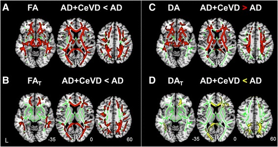
Free water (FW)-corrected measures revealed more lobar white matter (WM) tissue-related microstructural damage in patients with Alzheimer’s disease with cerebrovascular disease (AD + CeVD) than in patients with AD without CeVD. a On the basis of the conventional diffusion tensor imaging (DTI) model, patients with AD + CeVD exhibited reduced fractional anisotropy (FA) (red) compared with patients with AD in most of the WM regions, including the lobar and subcortical fibers. b Free water-corrected fractional anisotropy (FAT) revealed more lobar WM microstructural damage in patients with AD + CeVD than in patients with AD without CeVD, sparing the subcortical and brainstem regions. The WM skeleton is highlighted in green. c On the basis of the original DTI model, the patients with AD + CeVD had widespread increased axial diffusivity (DA) compared with patients with AD (red). d Following FW correction, a focal lobar (frontal and occipital lobes) free water-corrected axial diffusivity (DAT) reduction was identified in patients with AD + CeVD compared with patients with AD (yellow). The white matter skeleton is highlighted in green. Results are reported at p < 0.01, threshold-free cluster enhancement- and family-wise error-corrected