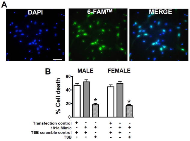
Figure 5. Cell death following miR-181a/ERα binding inhibition in astrocytes.
(A) Primary astrocytes stained with the nuclear dye DAPI (4′,6′-diamidino-2-phenylindole, blue, left panel) display 6-FAM™ reporter fluorescence (green, middle panel) when transfected with miR-181a/ERα target site blocker (TSB). Transfection efficiency is quantified by blue/green co-localization (right panel). Bars = 15 μm. (B) Quantification of cell death from GD injury of primary male and female astrocytes treated with control transfection or miR-181a mimic and TSB scramble control sequence or miR-181a/ESRα TSB. N = 8 cultures per experiment, mean±SE, * = p<0.05 versus control.

