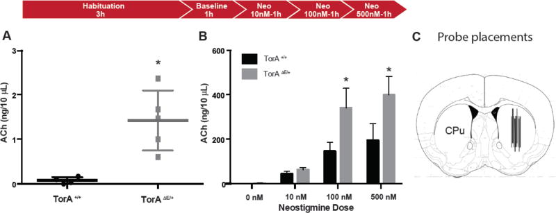
Figure 1. Basal ACh measured in vivo is elevated in DYT1 mouse model.
(A) TorAΔE /+ mice show siginificantly increased basal extracellular ACh levels; *p< 0.05. (B) Dose-related ACh increase in both TorA+/+ and TorAΔE /+ animals with neostigmine bromide in aCSF; *p< 0.05 ANOVA with Tukey’s post-hoc test for individual concentrations. (C) Diagram of probe placements in striatum (CPu).

