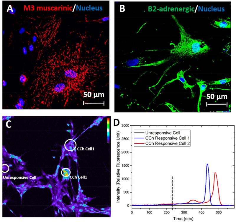
Figure 2. Functional Responses.
Expression and localization of M3 muscarinic (A, red) and β2-adrenergic (B, green) receptors and intracellular calcium signals in hSMECs (C, D). The [Ca2+i] signals were detected by treating hSMECs with M3 muscarinic acid agonist carbachol (CCh, 200 mM) and the response was observed by a subpopulation of hSMECs cultured on glass (C). The relative fluorescent intensity of [Ca2+i] in response to CCh over time was plotted for CCh-responsive (blue and red traces) and non-responsive (black trace) cells in D.

