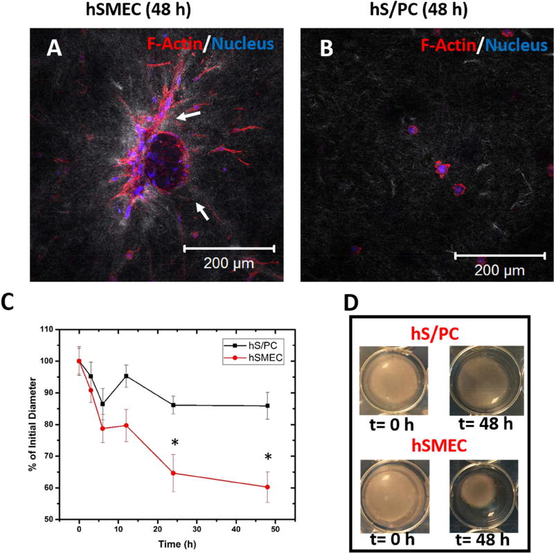
Figure 3. Collagen gel contraction assay.
(A-B). Differential reflectance confocal microscopy of hSMECs (A) and hS/PCs (B) cultured in collagen gels. F-actin and nuclei were stained red and blue, respectively, while collagen fibrils appeared grey (C-D). Reduction of the diameter of cell-laden collagen gels as a semi-quantitative assessment of hS/PCs and hSMECs contractility. The gross appearance of the gel constructs at 0 h and 48 h is shown in D. *: statistically significant when compared to hS/PC at the same time point, p<0.05

