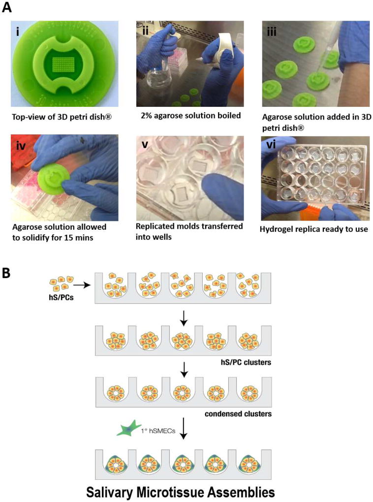
Figure 4. Step-wise assembly of salivary gland microtissues using agarose-replicated microwells.
(A) Photographs showing the hydrogel replication protocol. A commercial 3D petri dish® (i) was used as the mold. Sterilized molds were placed in a laminar flow hood and 2% agarose was brought to a boil (ii). The agarose solution was then added into the mold (iii). After the solution solidified (iv), the hydrogel replica was removed from the mold and transferred to a 24-well plate (v-vi) for cell assembly. (B) Schematic depiction of the bottom-up assembly process. Homogenously dispersed hS/PCs were counted and seeded in the microwell at a predetermined cell concentration for the targeted spheroid diameter. After an initial incubation period, cells aggregated and compacted into tight spheroid structures. Following spheroid formation, myoepithelial cells were added to the tight structures to form the microtissues.

