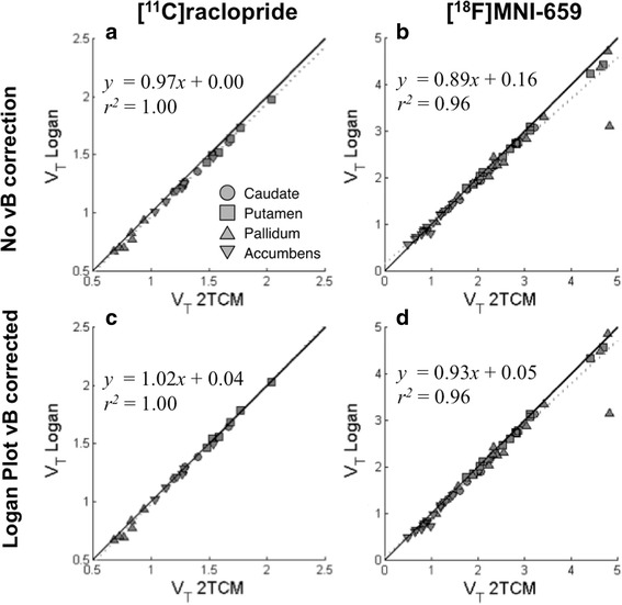
Fig. 3.

Distribution volumes (V T) obtained with 2TCM plotted against those obtained with Logan plot for all regions except cerebellum where V T from Logan plot are (a, b) not corrected, and (c, d) corrected for signal from blood vessels, for [11C]raclopride data (a, c) and [18F]MNI-659 data (b, d)
