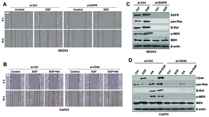
Figure 7.
Downregulation of EGFR and CD44 with siRNA inhibited the migration of ovarian cancer cells. (A) SK-OV-3 cells were treated with si-EGFR or si-Ctrl and EGF for 16 h. Migratory capabilities were analyzed using a wound healing assay. (B) Protein expression levels of Ras, Raf, p-MEK and total MEK were assessed using western blot analysis in SK-OV-3 cells. (C) Caov-3 cells were treated with si-CD44 or si-Ctrl and EGF or EGF combined with HA for 16 h. Migratory capabilities were analyzed using a wound healing assay. (D) Protein expression levels of Ras, Raf, p-MEK and total MEK were assessed using western blot analysis in Caov-3 cells. β-actin served as the loading control. Photomicrographs were captured at ×100 magnification using a digital camera under an inverted phase-contrast microscope. Scale bars, 100 µm. Results are representative of three independent experiments. EGF, epidermal growth factor; EGFR, EGF receptor; CD, cluster of differentiation; si, small interfering; Ctrl, control; p-, phosphorylated; MEK, mitogen-activated protein kinase/extracellular signal-regulated kinase kinase; HA, hyaluronan.

