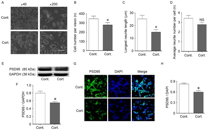
Figure 2.
Corticosteroid administration induced injury in N2a cells. (A-D) Cell morphological changes following corticosteroid administration at a dose of 2 µM for 48 h. (A and B) Corticosteroid treatment reduced the cell density. The cell number was reduced following treatment compared with the control cells. (A and C) Corticosteroid treatment shortened the neurite outgrowth length of the N2a cells, and the longest neurite length was significantly decreased compared with the control cells. (A and D) There was no significant difference in the average neurite number per cell. (E and F) The expression of synapse-associated protein PSD95 decreased. Western blotting indicated a decrease in PSD95 after 48 h of corticosteroid treatment at a dose of 2 µM. GAPDH was used as the loading control. (G and H) The location and decreased expression of PSD95 were demonstrated by immunofluorescence staining. DAPI was used to stain the nucleus. (G) PSD95 was located at the neurite outgrowth of N2a cells. (G and H) Corticosteroid treatment decreased the fluorescent intensity of PSD95 in N2a cells. For all panels, at least 100 cells in each culture were evaluated, and at least 3 cultures were used per group. The magnification and scale are as follows: (A) magnification, ×40; (G) magnification, ×200; bar, 20 µm. Data are presented as the mean ± standard error of the mean. *P<0.05 vs. control cells; NS, P>0.05 vs. control cells. N2a, neuro-2a; PSD95, postsynaptic density 95; cont, control cells; cort, corticosteroid treated cells.

