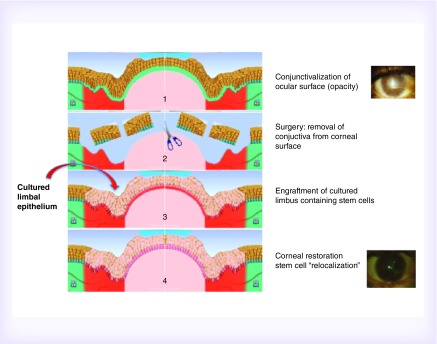
Figure 3. . Clinical application of cultured limbal stem cells.
The fibrovascular conjunctival pannus, grown on the corneal surface (panel 1), is removed (panel 2) to enable the transplant of the cultured limbal corneal epithelium (panel 3). Stem cell re-localization follows the cornea restoration (panel 4).

