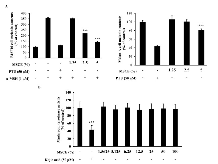
Figure 2.
Effects of MSCE treatment on melanogenesis in mouse melanocyte cells. (A) B16F10 mouse melanoma and melan-A mouse melanocyte cells were incubated with MSCE (1.25–5%) or PTU (50 µM) for 3 days, and melanin content was measured. B16F10 cells were additionally induced towards melanogenesis with 1 µM α-MSH. (B) Mushroom TYR activity was measured in the presence of MSCE (0–100%). Kojic acid, a known inhibitor of TYR activity, was used as a positive control. Each experiment was conducted in triplicate and the data are presented as the mean ± standard deviation. ***P<0.001 vs. α-MSH-treated B16F10 cells or untreated Melan-A cells. α-MSH, α-melanocyte stimulating hormone; MSCE, mixed Stichopus japonicus extract; PTU, phenylthiourea; TYR, tyrosinase.

