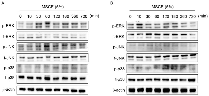
Figure 4.
Effects of MSCE on signal transduction pathways in mouse melanocyte cells. Following 24 h of serum starvation, (A) B16F10 mouse melanoma cells and (B) Melan-A mouse normal melanocytes were treated with 5% MSCE for the indicated time periods. Cell lysates were harvested for western blot analysis using primary antibodies against p-ERK, t-ERK, p-JNK, t-JNK, p-p38 and t-p38; β-actin was used as the loading control. The results are representative of triplicate experiments. α-MSH, α-melanocyte stimulating hormone; ERK, extracellular signal-regulated kinase; JNK, c-Jun N-terminal kinase; MSCE, mixed Stichopus japonicus extract; p, phosphorylated; p38, mitogen-activated protein kinase 14; t, total.

