Figure 2.
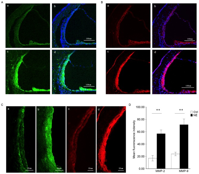
Immunofluorescence assessment of MMP-2 and MMP-9 expression in the stria vascularis prior to and following NE. (A) Tissue double-labeled for MMP-2 (green) and DAPI (blue) prior to (a, b) and following (c, d) noise exposure. Scale bar, 100 µm. (B) Tissue double-labeled for MMP-9 (red) and DAPI (blue) prior to (a, b) and following (c, d) noise exposure. Scale bar, 100 µm. (C) Weak MMP-2 (green) and −9 (red) immunoreactivity was observed in marginal cells and basal cells in the stria vascularis of controls (a, c), which significantly increased following noise-trauma (b, d). Scale bar, 25 µm. (D) Quantification of localized fluorescence density of MMP-2 (green) and −9 (red) in the stria vascularis. Data are expressed as the mean ± standard deviation. **P<0.01. MMP, matrix metalloproteinase; NE, noise exposure; Ctrl, control.

