Figure 3.
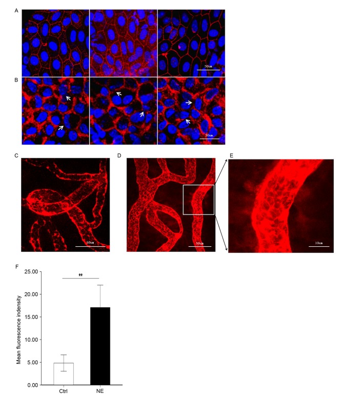
Noise trauma upregulates expression of ZO-1 (red) and causes alterations in BLB permeability, nuclei are labeled by DAPI (blue). Magnification, ×60. (A) Representative confocal microscope images demonstrating the compact linear structure of ZO-1 (red) on the plasma membrane in the stria vascularis. Scale bar, 30 µm. (B) Noise exposure caused the structure of ZO-1 (red) to loosen, and regional breaks were observed (white arrows). Scale bar, 30 µm. BLB integrity was assessed by noting the degree of EBD extravasated around strial capillaries (C) prior to and (D) following noise exposure. Scale bar, 50 µm. (E) EBD outside capillaries was observed further away from the vessel with noise exposure, demonstrative of the significantly increased permeability following loud noise exposure. Scale bar, 10 µm. (F) Mean fluorescence density outside capillaries in the stria vascularis, as assessed with Image J software. Data are expressed as the mean ± standard deviation. **P<0.01. EBD, Evans blue dye; ZO-1, zona-occludens 1; Ctrl, control; NE, noise exposure; Ctrl, control.

