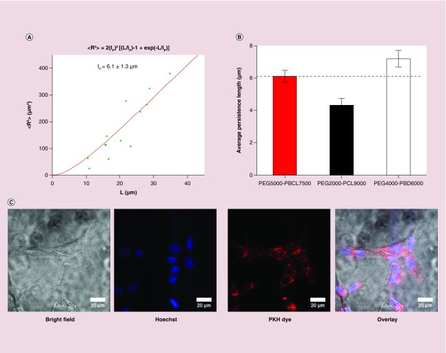
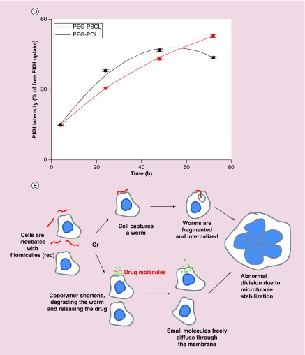
Figure 2. . Cellular uptake of filomicelles.
(A) Model fitting during the calculation of persistence length of PEG–PBCL filomicelles. (B) While the persistence length of PEG–PBCL is higher than that of PEG–PCL, it is lower than PEG–PBD (also proven to form flexible filomicelles). (C) PKH, Hoechst and bright field images of cells incubated with PKH-labeled filomicelles. Cytoplasmic spots of the dye after filomicelle uptake by EC4 cells can be seen after incubation for one day. Scale bars are 20 μm. (D) Quantification of PKH intensity within a cell after incubation with dye-labeled filomicelles. The intensity recorded increases exponentially with time for PEG–PBCL, while it is parabolic for PEG–PCL worms. PEG–PCL worms have higher accumulation initially, but is surpassed by PEG–PBCL on day 3. (E) Schematics depicting the uptake of filomicelles (or worms) by cells. Cells, when incubated with filomicelles, come in contact with and capture them. The cell may then proceed to chew off a part of the filomicelle. Alternatively, the constituent diblock copolymers may undergo hydrolysis leading to its shortening. The corresponding phase transition of filomicelles to spheres destabilizes the filomicelles, leading to release of the encapsulated drugs. These small molecules are then taken up by the cell.


