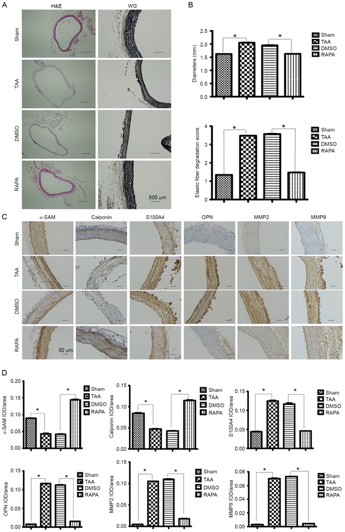
Figure 2.
Expression of contractile proteins and dedifferentiation markers in aortic segments 2 weeks post-rapamycin treatment. (A) Representative lower-power micrographs of the hematoxylin and eosin and Verhoeff-Van Gieson staining of NaCl-treated (sham group, n=6) and CaCl2-treated aortas (TAA group, n=6) pretreated with rapamycin (RAPA group, n=8) and DMSO (DMSO group, n=8). Scale bar, 500 µm. (B) Measurements of external media diameter and grading of elastin degradation in the four groups (2 weeks post-TAA induction). Data are presented as the mean ± standard error of mean, *P<0.05 as indicated. (C) Representative pictures of immunohistochemical study in aortic segments 2 weeks post-TAA induction. The slides were stained with αSMA, calponin, S100A4, OPN, MMP2 and MMP9. The anti-rabbit horseradish peroxidase/diaminobenzidine detection system was used to visualize the expression (brown staining). Scale bar, 50 µm. (D) The quantitation of the protein content of αSMA, calponin, S100A4, OPN, MMP2 and MMP9. The immunohistologically stained slides were assessed by computerized planimetry in the aortic adventitia and aortic media. Data are presented as the mean ± standard error of mean. *P<0.05 as indicated. TAA, thoracic aortic aneurysm; RAPA, rapamycin; MMP, matrix metallopeptidase; OPN, osteopontin; αSMA, α-smooth muscle actin; IOD, integral optical density.

