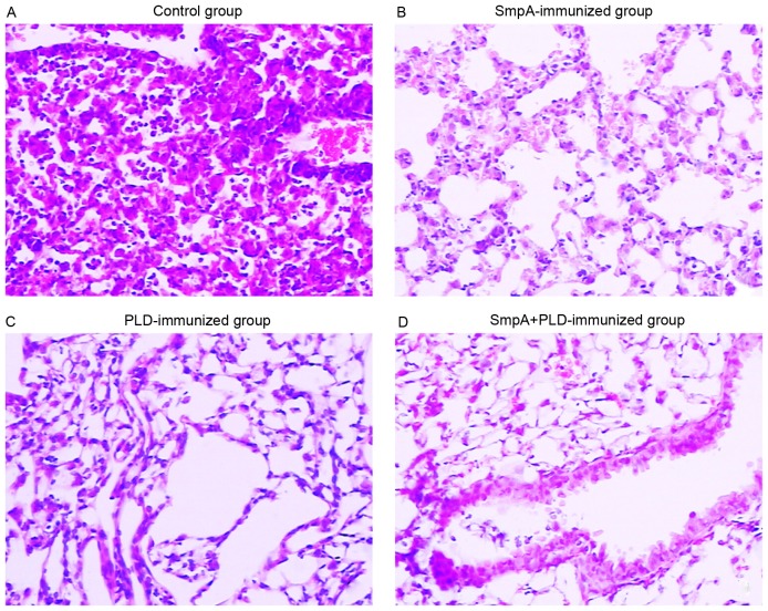
Figure 6.
Photomicrograph images of lung histopathology. Photomicrograph images (magnification, ×200; hematoxylin and eosin staining) of lung histopathology in (A) control mice, (B) SmpA-immunized mice, (C) PLD-immunized mice and (D) mice immunized with both SmpA and PLD. SmpA, small protein A; PLD, phospholipase D.

