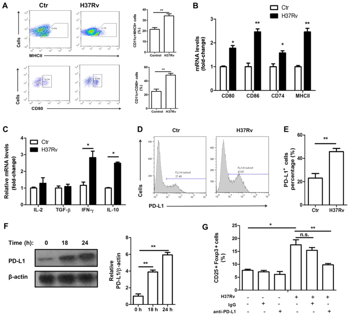
Figure 3.
H37Rv promotes macrophage activation and PD-L1 expression. (A) Macrophage activation markers MHCII and CD80 were tested by FACS analysis after H37Rv stimulation. (B) mRNA levels of CD80, CD86, MHCII and CD74 were determined by RT-PCR. (C) mRNA levels of IL-2, TGF-β, IFN-γ and IL-10 were determined by RT-PCR. (D and E) The surface expression of PD-L1 was analyzed by FACS. (F) Immunoblot analysis of total PD-L1 protein levels after H37Rv expression. (G) T cells were co-cultured with H37Rv-infected BMDMs in the presence of neutralizing antibody anti-PD-L1. CD25+Foxp3+ Tregs gated on CD4 were analyzed by flow cytometry. All experiments were representative of three similar results. Statistical differences between groups are indicated by the P-values. *P<0.05; **P<0.01.

