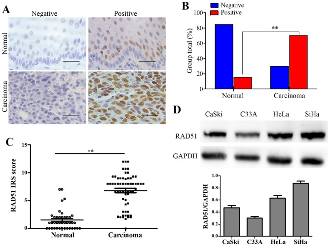
Figure 1.
RAD51 expression in normal cervix and cervical carcinoma tissues. (A) Immunohistochemistry results showing RAD51 expression in normal cervix and cervical carcinoma tissues; scale bar, 50 µm. (B) RAD51 staining is classified into negative and positive, and the percentage of tissues in each group is shown. (C) The IHC scores of RAD51 staining in the normal cervix and carcinoma tissues are shown. (D) The expression of RAD51 in Caski, C33-A, HeLa and SiHa cells was measured by western blotting, the relative expression of RAD51 was calculated based on western blot analyses, GAPDH was used as an internal control. *P<0.05, **P<0.01.

