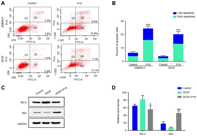
Figure 6.
P18 peptide induces apoptosis of HUVECs in vitro. (A) HUVECs were cultured with or without VEGF and the P18 peptide (0.32 nM) for 24 h. Apoptosis levels were evaluated by a flow cytometry assay. (B) Data represent the percentages of early and late apoptotic cells and are expressed as the mean ± SD, *P<0.05, ***P<0.01. (C) Expression levels of Bcl-2 and Bax in HUVECs were analysed by western blotting after culture with or without VEGF (8 ng/ml) and the P18 peptide (0.32 nM) for 24 h. (D) The density of each band in western blot assay was quantified and normalized to that of GAPDH (mean ± SD, *P<0.05, ***P<0.01).

