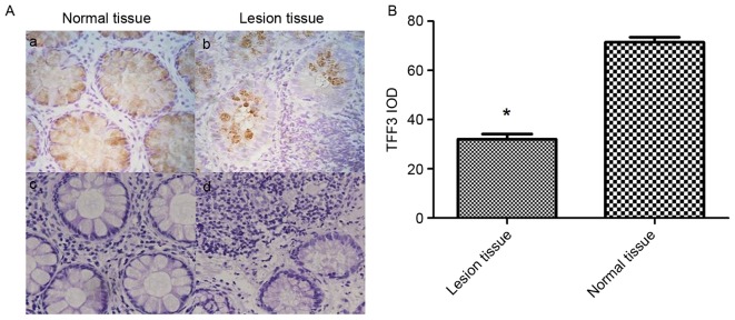
Figure 4.
TFF3 expression in lesional tissue from patients with IBD and normal control tissues. (A) Immunohistochemical staining was performed to determine the relative protein expression of TFF3 in IBD lesion colon tissues and normal control tissues (n=8; magnification, ×400). Immunohistochemical staining of (a) normal colonic tissue and (b) lesional colonic tissue; H&E staining of (c) normal colonic tissue and (d) lesional colonic tissue. (B) IOD of TFF3 staining. *P<0.05 vs. normal tissue. H&E, hematoxylin and eosin staining; IBD, inflammatory bowel disease; IOD, integrated optical density; TFF3, trefoil factor 3.

